Click here to see the online version.
 |
Training on Auto Brightness and Contrast Control Imaging Techniques for Practical Bio-Medical Applications Klang Valley, Malaysia |
||||
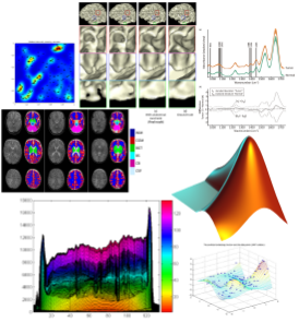
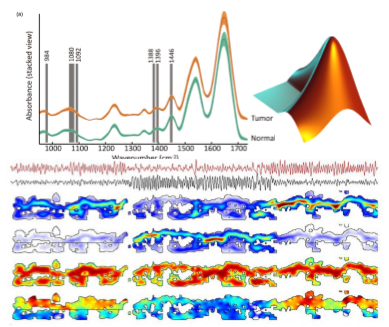
Image techniques for Bio-Medical has become a major aspect of engineering sciences and radiology in particular has become a dominant player in the field. Recent development have made it possible to use biomedical imaging to view the human body from an anatomical or physiological prospective in a non-invasive fashion. By the increasing use of direct digital imaging
systems for medical diagnostics, digital image
processing becomes more and more important in health care. In additional to originally digital
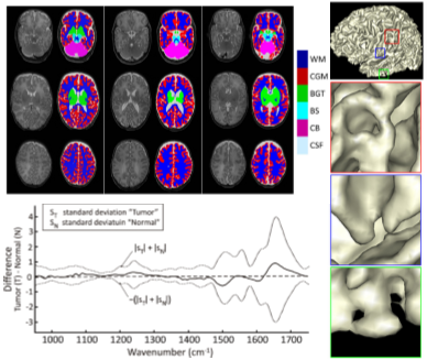
methods. Such as Computed Tomography (CT) or Magnetic Resonance Imaging (MRI), initially analogue imaging modalities such as endoscopy or radiography are nowadays equipped with digital sensors. Digital images are composed of individual pixels to which discrete brightness or color values are assigned. They can be efficiently processed, objectively evaluated, and made available at many places at the time by means of appropriate communication networks such as PACS and DICOM protocol. Based on digital
imaging techniques, the entire spectrum of digital image processing is now applicable in medicine. |
 |
|
 |
Course Objectives Course Methodology |
|
Course highlights Course Agenda Fundamental of Medical Imaging? |
Practical and application specific exercises, and case studies All cases studies will cover the basic practice by medical doctors, spatial based Prerequisites |
|
|
Who should attend?
Scientists, mathematicians, engineers and programmers at all levels who work with or need to learn about fundamental of biomedical Imaging concepts and techniques. No background in either of these topics are, however, assumed. The detailed course material and many source code listings will be invaluable for both learning and reference.
Sign Up Now!
Other upcoming courses to enrich and enhance your technical computing capability and data anaysls skill-set, Just click onto the Title to find out more details.
 |
 |
 |
 |
 |
 |
 |
 |
 |
 |
 |
 |
For more information, please contact us at enquiry@solutions4u-asia.com
The updates of our training program are sent to you, as we think that they might be of interest and benefit you. Please help to forward to others who may be interested. However, you may unsubscribe if you do not wish to receive further mailing from us. Thank you.
